-
Author
Mauranda Men -
PI
Dr. Edmund Tsui
-
Co-Author
Nina Cherian
-
Title
Hyper-Parallel Optical Coherence Tomography Imaging of Herpes Zoster Keratitis
-
Program
STTP
-
Other Program (if not listed above)
-
Abstract
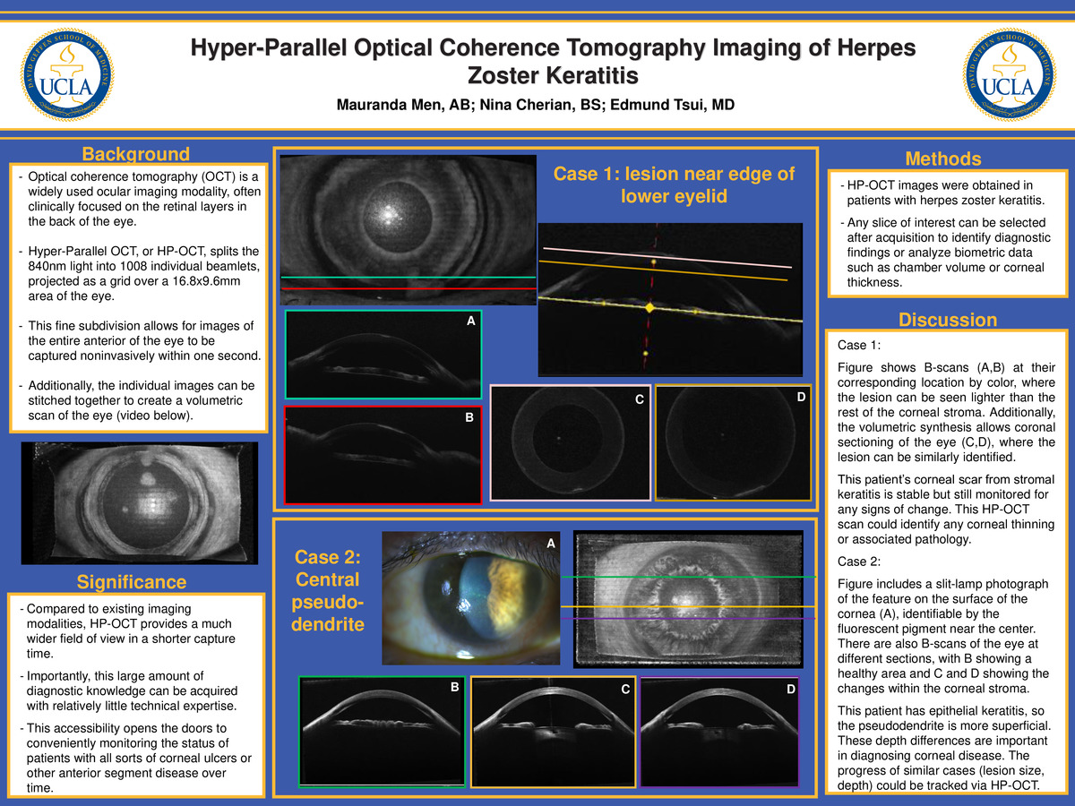
Optical coherence tomography (OCT) is a widely used ocular imaging modality, which uses light wavelengths to obtain cross-sectional images of the eye in a method mechanically similar to ultrasound. Clinically, OCT is most often focused on the retinal layers in the back of the eye, although it can also be diagnostically helpful in distinguishing lesions and pathologies in the anterior segment. This investigation demonstrates the capabilities of Hyper-Parallel OCT (HP-OCT), a newly developed imaging platform, on patients with herpes zoster ophthalmicus (HZO) who have stromal scarring, epithelial keratitis, and stromal keratitis. Unlike conventional OCT technology, the HP-OCT machine splits the light source into a grid of individual beamlets that are analyzed in parallel, allowing for image acquisition of the entire front of the eye (16.8mm x 9.6mm) in under one second. Each individual image is then stitched together into a volumetric scan of the eye, allowing for the user to view a sliced scan of the eye at any angle and from any position. After the images are acquired and analyzed, any section can then be used to calculate biometric data, from corneal thickness to anterior chamber volume. This rapid image capture time allows for a large amount of diagnostic data to be captured quickly, with relatively little technical expertise required. As accessibility widens, future development of this new imaging modality may allow for quantitative evaluation in the monitoring of corneal inflammatory diseases.
-
PDF
-
Zoom
https://uclahs.zoom.us/j/96170775186?pwd=QmFqTm45ak9UNzR5T2VGdWJIT2d6UT09
