-
Author
Eghosa Edogun -
PI
Kristofer Jones
-
Co-Author
Eghosa Edogun, William Sheppard
-
Title
Surgical Fixation of Chondral-Only Fragments: A Systematic Review
-
Program
CTSI TL1 Summer Program
-
Other Program (if not listed above)
-
Abstract
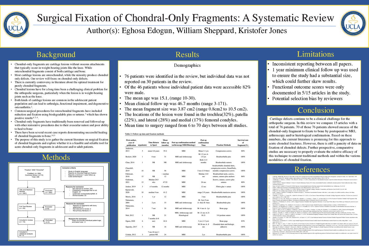
Introduction Chondral fragments are cartilage lesions without osseous attachments that typically occur in weight-bearing joints like the knee. There is currently a controversy in the literature about the optimal treatment for these purely chondral fragments.
Background Altogether cartilage lesions continue to present as challenging clinical problems for the orthopedic surgeon. These injuries typically occur due to osteochondritis dissecans, patellar dislocation, or trauma9. Common surgical procedures for osteochondral fragments have been reduction and fixation using biodegradable pins or sutures 3. This method for fixation of osteochondral lesions has shown to have a high rate of bone-to-bone healing and good clinical outcomes. 4,5,6 However, fragments without bony attachments have traditionally been removed because of their avascular nature and poor ability to heal to bone 7.
Methods: A thorough search of the literature on fixation of chondral fragments was done and reviewed in conjunction with PRISMA (Preferred Reporting Items for Systematic Reviews and Meta-Analyses).
Results 15 studies were initially screened, and information about 76 patient knees was extracted and compared. Of the 46 patients whose individual patient data was accessible, 82% were male. The mean age was 15.1 (range10-30). Mean clinical follow-up was 46.7 months (range 3-171). The mean fragment size was 3.87 cm2 (range 0.8cm2 to 10.5 cm2). The locations of the lesion were in the trochlea(32%), patella (22%), and lateral (28%), and medial (17%) femoral condyles. Meantime to surgery ranged from 6 to 70 days between all studies. The mean clinical follow-up ranged from 12-61.2 months between all patients. 70 of those 76 patients experienced success of the fragment to fixate to the bone by Postoperative MRI, arthroscopy, and histological confirmation.
Conclusion The current literature is promising for the utilization of surgical fixation of chondral fragments. Further prospective, comparative studies are necessary to properly evaluate the relative efficacy of this approach to current traditional methods.
-
PDF
-
Zoom
https://uclahs.zoom.us/j/95040499795?pwd=cTF4a0hSUXBxQVhTREMxTi9CV3p5QT09
