-
Author
Nelly Kokikian -
PI
Jonathan Wanagat, M.D., Ph.D.
-
Co-Author
Amy Vandiver, M.D., Ph.D.
-
Title
Optimization of staining and quantifying mitochondrial deletions in photo-aged skin
-
Program
Medical Student Training in Aging (MSTAR) Research Program
-
Other Program (if not listed above)
-
Abstract
Title: Optimization of staining and quantifying mitochondrial deletions in photo-aged skin
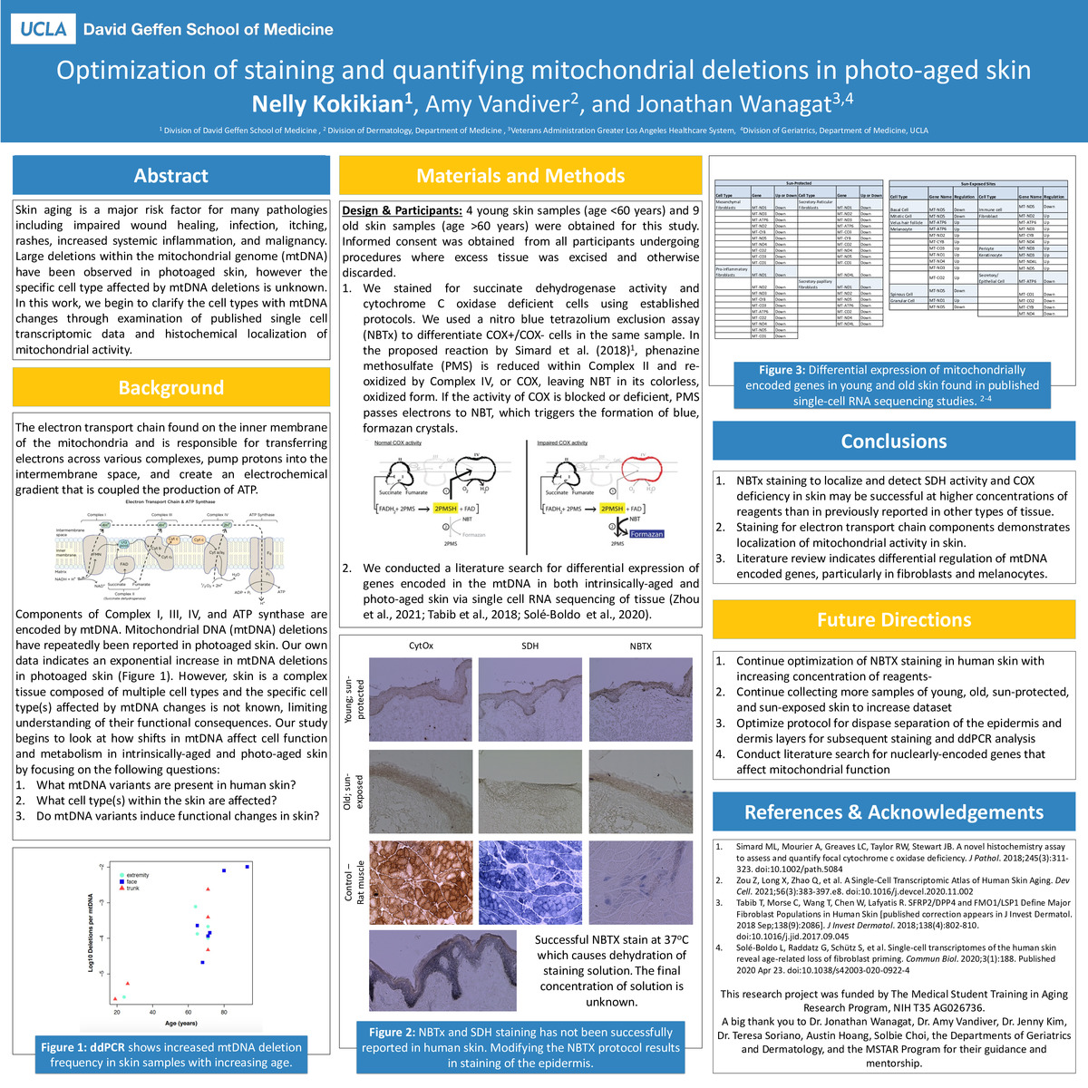
Importance: Skin aging is a major risk factor for many pathologies including impaired wound healing, infection, itching, rashes, increased systemic inflammation, and malignancy. Large deletions within the mitochondrial genome (mtDNA) have been observed in photoaged skin, however the specific cell type affected by mtDNA deletions is unknown. In this work, we begin to clarify the cell types with mtDNA changes through examination of published single cell transcriptomic data and histochemical localization of mitochondrial activity.
Objective: To determine how shifts in mtDNA affect cell function and metabolism in intrinsically-aged and photo-aged skin by focusing on the following questions:
- What mtDNA variants are present in human skin?
- What cell type(s) within the skin are affected?
- Do mtDNA variants induce functional changes in skin?
Design & Participants: 4 young human skin samples (age < 60 years) and 9 old human skin samples (age > 60 years) were obtained for this study. Samples were stained for succinate dehydrogenase activity and cytochrome C oxidase deficiency using the nitro blue tetrazolium exclusion assay (NBTx). A literature search was conducted identify differentially expressed of genes encoded in the mtDNA in both intrinsically-aged and photo-aged skin via single cell RNA sequencing of tissue.
Main Outcome(s) and Measure(s):
- To optimize NBTx staining to detect SDH staining and COX deficiency in human skin to localize mitochondrial activity in skin.
- To determine which cell types have differentially expressed mitochondrially-encoded genes.
Results: Multiple iterations of NBTx staining using standard protocol concentrations in young, sun-protected, old, and sun-exposed tissue samples were mostly unsuccessful except for one sample whose staining solution became more concentrated during incubation. Literature review indicates differential regulation of mtDNA encoded genes, particularly in fibroblasts and melanocytes.
Conclusions and Relevance: NBTx staining in human skin to localize and detect SDH activity and COX deficiency in skin may be successful at higher concentrations of reagents than in previously reported in other types of tissue. Further optimization of the staining protocol will allow for localization COX+/COX- cells and mitochondrial activity in skin. Skin is a complex tissue composed of multiple cell types and the specific cell type(s) affected by mtDNA changes are not known, limiting understanding of their functional consequences. MtDNA contains genes that encode components of Complex I, III, IV, and ATP synthase and are responsible for energy production in cells. Investigating mtDNA deletions and resulting metabolic changes in mitochondria in skin will clarify the role of mtDNA deletions on shifts in mitochondrial metabolism and function.
-
PDF
-
Zoom
https://uclahs.zoom.us/j/92932433046?pwd=WHNyNkh2eEpSL3NuVXExenN5NndVQT09

