-
Author
Connie Ho -
PI
Joseph Caprioli, MD
-
Co-Author
Victoria L. Tseng, MD, PhD, Esteban Morales, MS, Fei Yu, PhD, Anne L. Coleman, MD, PhD, Joseph Caprioli, MD
-
Title
Phenotypic Subtypes of Glaucoma Patients with Small & Large Optic Discs
-
Program
STTP
-
Other Program (if not listed above)
-
Abstract
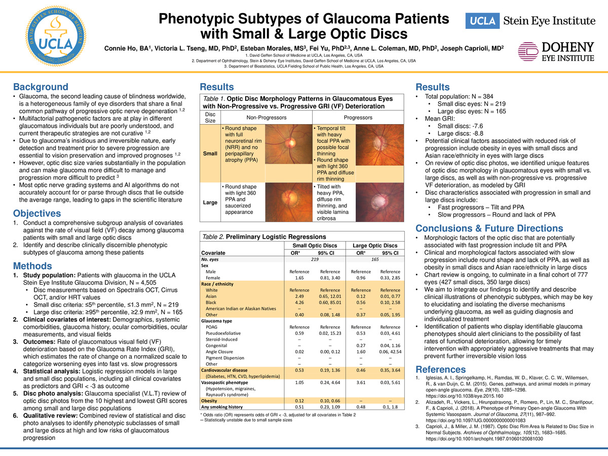
Background: Glaucoma, the second leading cause of blindness worldwide, is a heterogeneous family of eye disorders that share a final common pathway of progressive optic nerve degeneration. Multifactorial pathogenetic factors are at play in different glaucomatous individuals but are poorly understood, and current therapeutic strategies are not curative. Due to glaucoma’s insidious and irreversible nature, early detection and treatment prior to severe progression are essential to vision preservation and improved prognoses. However, optic disc size varies substantially in the population and can make glaucoma more difficult to manage and progression more difficult to predict. Most optic nerve grading systems and AI algorithms do not accurately account for or parse through discs that lie outside the average “normal” range, leading to gaps in the scientific literature.
Objectives: To identify and describe clinically discernible phenotypic subtypes of glaucoma among patients with small and large discs, by conducting a comprehensive subgroup analysis of covariates against the rate of visual field (VF) decay among glaucoma patients with small and large optic discs, and by reviewing their corresponding images.
Methods: In this retrospective study, the study population included all patients with glaucoma in the UCLA Stein Eye Institute Glaucoma Division (N = 4,505). Optic disc measurements based on Spectralis OCT, Cirrus OCT, and/or HRT values were used to identify eyes with smaller and larger discs than normal (small disc criteria: ≤5th percentile, ≤1.3 mm2, N = 219; large disc criteria: ≥95th percentile, ≥2.9 mm2, N = 165). Patient charts were reviewed for clinical covariates of interest, including: demographics, systemic comorbidities, glaucoma history, ocular comorbidities, ocular measurements, and visual fields. Glaucomatous visual field (VF) rates of deterioration were measured based on the Glaucoma Rate Index (GRI), which estimates the rate of change on a normalized scale to categorize worsening eyes into fast vs. slow progressors. Logistic regression models were performed in the small and large disc populations, including all clinical covariates as predictors and GRI < -3 as the outcome. Stereoscopic disc photos of the 10 highest and lowest GRI scores for small and large disc populations were reviewed by a glaucoma specialist (VLT). Qualitative review of statistical results and disc photo analyses were combined to identify phenotypic subclasses of small and large discs at high and low risk of glaucomatous progression.
Results: The total study population included 384 eyes, of which 219 had small discs and 165 had large discs. Mean GRI for the small discs was -7.6, and mean GRI for the large discs was -8.8. In logistic regression models, factors associated with decreased progression included obesity in small discs (adjusted odds ratio [aOR] = 0.12, 95% confidence interval [CI] = 0.10, 0.66) and Asian race/ethnicity in large discs (aOR = 0.12, 95% CI = 0.01, 0.77). On review of disc photos, disc characteristics associated with progression in small and large discs include tilt and peripapillary atrophy (PPA) for fast progressors, and round disc shape and lack of PPA for slow progressors.
Conclusions & Future Directions: In our preliminary analyses, clinical and morphologic factors potentially associated with fast progression include tilt and PPA. Factors potentially associated with slow progression include round disc shape and lack of PPA, as well as obesity in small discs and Asian race/ethnicity in large discs. Chart review is ongoing, to culminate in a final cohort of 777 eyes (427 small discs, 350 large discs). We aim to integrate our findings to identify and describe clinical illustrations of phenotypic subtypes, which may be key to elucidating and isolating the diverse mechanisms underlying glaucoma, as well as guiding diagnosis and individualized treatment. Identification of patients who display identifiable glaucoma phenotypes should alert clinicians to the possibility of fast rates of functional deterioration, allowing for timely intervention with appropriately aggressive treatments that may prevent further irreversible vision loss.
-
PDF
-
Zoom
https://uclahs.zoom.us/j/95054923270?pwd=UEtEdm9ST2RDRVkyYXY3MFdjUStlQT09
