-
Author
Simon Levinson -
Co-author
Michelle Miller, Ahmed Iftekhar, Monica Justo, Daniel Arriola, Samman Hazany, Josue Avecillas-Chasin, Ausaf Bari
-
Title
A Structural Connectivity Atlas of Limbic Brainstem Nuclei
-
Abstract
Background
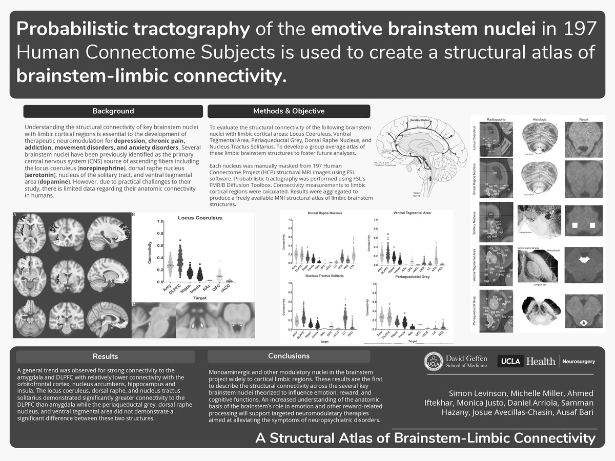
Understanding the structural connectivity of key brainstem nuclei with limbic cortical regions is essential to the development of therapeutic neuromodulation for depression, chronic pain, addiction, movement disorders, and anxiety disorders. Several brainstem nuclei have been previously identified as the primary central nervous system (CNS) source of ascending fibers including the locus coeruleus (norepinephrine), dorsal raphe nucleus (serotonin), nucleus of the solitary tract, and ventral tegmental area (dopamine). However, due to practical challenges to their study, there is limited data regarding their anatomic connectivity in humans.
Objective
To evaluate the structural connectivity of the following brainstem nuclei with limbic cortical areas: Locus Coeruleus, Ventral Tegmental Area, Periaqueductal Grey, Dorsal Raphe Nucleus, and Nucleus Tractus Solitarius. To develop a group average atlas of these limbic brainstem structures to foster future analyses.
Methods
Each nucleus was manually masked from 197 Human Connectome Project (HCP) structural MRI images using FSL software. Probabilistic tractography was performed using FSL’s FMRIB Diffusion Toolbox. Connectivity measurements to limbic cortical regions were calculated. Results were aggregated to produce a freely available MNI structural atlas of limbic brainstem structures.
Results
A general trend was observed for strong connectivity to the amygdala and DLPFC with relatively lower connectivity with the orbitofrontal cortex, nucleus accumbens, hippocampus and insula. The locus coeruleus, dorsal raphe, and nucleus tractus solitarius demonstrated significantly greater connectivity to the DLPFC than amygdala while the periaqueductal grey, dorsal raphe nucleus, and ventral tegmental area did not demonstrate a significant difference between these two structures.Conclusion
Monoaminergic and other modulatory nuclei in the brainstem project widely to cortical limbic regions. These results are the first to describe the structural connectivity across the several key brainstem nuclei theorized to influence emotion, reward, and cognitive functions. An increased understanding of the anatomic basis of the brainstem’s role in emotion and other reward-related processing will support targeted neuromodulatary therapies aimed at alleviating the symptoms of neuropsychiatric disorders.
-
College
AAC
-
Zoom
https://ucla.zoom.us/j/99212331237?pwd=dVMzOG9KTmdVcVNUajVCZHVDdjFpZz09
-
PDF