-
Author
Angela Chen -
Co-author
Nathaniel P. Pelsor, Kaidi Wang, Ben J. Glasgow, Anthony J. Aldave
-
Title
Late Onset Interface Calcium Deposition Following LASIK
-
Abstract
Purpose: To report a novel clinical entity characterized by bilateral calcium deposits in the flap interface following uncomplicated laser in situ keratomileusis (LASIK).
Methods: Slit lamp examination, anterior segment optical coherence tomography (AS-OCT) imaging and histopathologic analysis of an interface opacity were performed to characterize and identify the origin of the interface opacities.
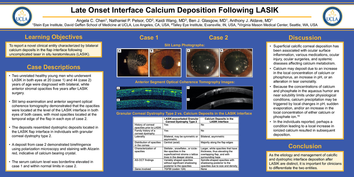
Results: Two unrelated healthy young men who underwent LASIK in both eyes at 20 (case 1) and 44 (case 2) years of age were diagnosed with bilateral, white anterior stromal opacities five years after LASIK surgery. Slit lamp examination and AS-OCT imaging demonstrated that the opacities were located at the level of the LASIK interface in both eyes of both cases, with the majority of the opacities located at the temporal edge of the flap in each eye of case 2. An opacity from case 2 demonstrated birefringence using polarization microscopy and staining with Alizarin red, indicative of calcium bearing crystal. The serum calcium level was borderline elevated in case 1 and within normal limits in case 2.
Conclusions: Intrastromal calcium deposition can occur after LASIK surgery, with the deposits resembling dystrophic deposits located in the LASIK flap interface in individuals with granular corneal dystrophy type 2. As the etiology and management of calcific and dystrophic interface deposition after LASIK are distinct, it is important for clinicians to differentiate the two entities based on the examination, diagnostic imaging and if necessary, molecular genetic analysis.
-
College
AAC
-
Zoom
-
PDF